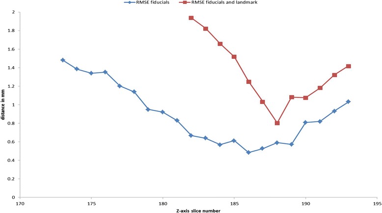
Fig. 6.

Identification of corresponding CT planes and histology slices. Example showing how corresponding histology slices and (ex vivo) CT planes are identified. The RMS distance is calculated using either four spine fiducial markers (blue line) or four spine fiducial markers and one anatomical landmark (red line). Although there is approximate agreement between the two methods, the use of landmarks leads to more precise identification as the landmarks are often confined to one or two consecutive axial slices. Although the measurements using the fiducial markers only (blue line) leads to smaller RMSE, both methods lead to a very similar position of the global minimum. The CT slice that showed corresponding to the minimum RMSE was chosen as that corresponding with the given histopathology slice. RMSE root mean square error
