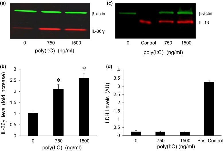
Fig. 1.
Low doses of poly(I:C) induce IL-36γ expression. a Representative western blot showing IL-36γ expression in keratinocytes stimulated with increasing concentrations of poly(I:C) for 48 h. b Quantification of intracellular IL-36γ. Bars show mean ± SD, relative to controls treated with vehicle (n = 6 experiments, *p < 0.001). c Representative western blot of keratinocytes treated with vehicle or with poly(I:C) for 48 h. Recombinant IL-1β at 20 ng/lane was used as a positive control. β-actin was used as a loading control. d Cells were treated with increasing concentrations of poly(I:C) for 96 h and the culture medium assayed for released lactic dehydrogenase (LDH) as a measure of cell death. Culture medium from cells lysed with the detergent NP40 served as a positive control

