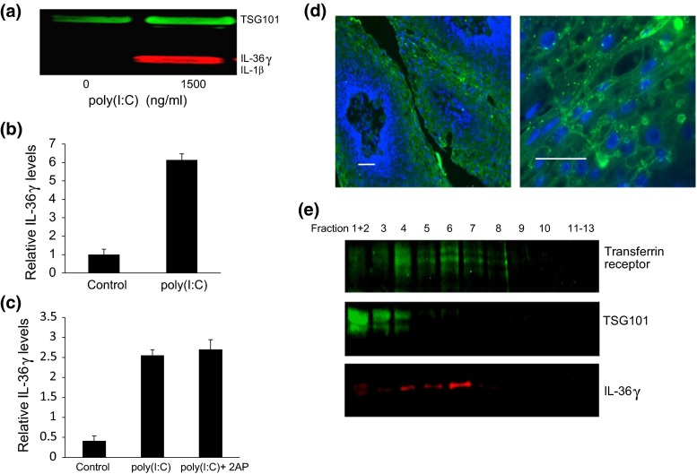
Fig. 5.
Poly(I:C) induces IL-36γ release in multiple extracellular vesicles (EVs), consistent with punctuate appearance of IL-36γ in suprabasal layers of papillomas. a Representative western blot. HFKs were stimulated with 1500 ng/ml poly(I:C) for 96 h, vesicles isolated by differential centrifugation, and analyzed by western blot using TSG101 as a marker for EVs and as a loading control. b Quantification of IL-36γ within vesicles. Bars show mean ± SD, relative to controls without poly(I:C) (n = 4 experiments, *p < 0.001). c Cells were treated with poly(I:C) ± 1 mM 2AP, and EVs isolated and analyzed as in “a.” Bars show mean ± SD, relative to controls of 4 experiments. d Sections of paraffin-imbedded papilloma tissues were incubated with goat anti-IL-36γ and visualized with fluorescein-conjugated donkey anti-goat IgG. DAPI staining of DNA was used as a counterstain. Images show representative papilloma sections. Bars = 40 µm. e Cells were stimulated with 1500 ng/ml poly(I:C) for 96 h, extracellular vesicles were extracted by differential centrifugation, separated on sucrose gradients and fractions from the gradients analyzed by western blot. TSG101 is a marker for endosomes and the transferrin receptor marks exosomes. A representative blot is shown

