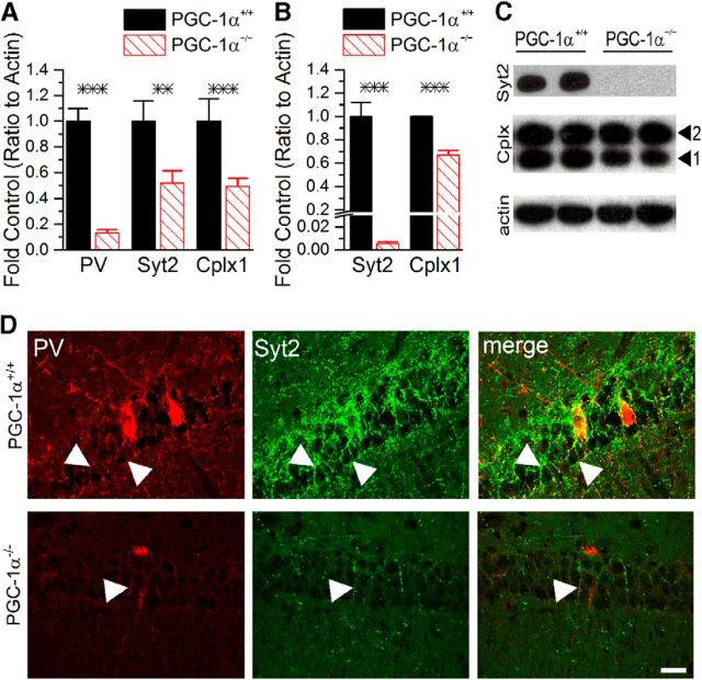
Figure 4.
PGC-1α is required for the expression of synaptic vesicle fusion proteins in hippocampus. A, q-RT-PCR revealed significant reductions in transcript expression of parvalbumin (PV), synaptotagmin 2 (Syt2), and complexin 1 (Cplx1) in hippocampal homogenates from PGC-1α−/− (n = 6–9 per group) compared with PGC-1α+/+ mice (n = 7–12 per group). B, Western blot analysis confirmed that protein expression of Syt2 and Cplx1 was also reduced in hippocampal homogenates from PGC-1α−/− (n = 7) compared with PGC-1α+/+ mice (n = 7). C, Representative Western blots of data shown in B. Arrowheads indicate bands for Cplx1 and Cplx2. D, Immunofluorescence double-labeling with antibodies against Syt2 (green) and PV (red) revealed high coexpression (yellow) of the two proteins in axon terminals throughout the CA1 pyramidal layer in PGC-1α+/+ mice. Syt2 is highly expressed in rings of PV-positive puncta (arrowheads) surrounding CA1 pyramidal cells. Both Syt2 and PV expression were markedly reduced in PGC-1α−/− mice, but limited coexpression in axon terminals was still evident (arrowhead). Scale bar, 25 μm. n = 3–5 per group for immunofluorescence experiments. ***p < 0.0005, **p < 0.005, two-tailed, independent-samples t tests.

