Figure 6.

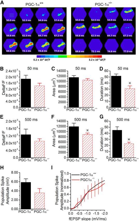
Partial restoration of circuit function at short paired-pulse intervals in PGC-1α−/− mice. A, Example images of the spatiotemporal pattern of voltage-sensitive dye signals evoked in CA1 by Schaffer collateral stimulation in slices from PGC-1α+/+ and PGC-1α−/− mice from the second pulse in a paired-pulse stimulation at a 50 ms interval. B, The maximum amplitude of the voltage-sensitive dye response is similar on the second pulse (n = 12, 7). C, The spread of the response is unchanged (n = 12, 7). D, The duration of the response is reduced at the 50 ms interval on the second pulse in PGC-1α−/− mice (n = 12, 7). E, The maximum amplitude of the voltage-sensitive dye response is not significantly different on the second pulse of a 500 ms interval (n = 11, 7). F, The spread of the response is decreased at the 500 ms interval (n = 11, 7). G, The duration of the response is decreased at the 500 ms interval in the PGC-1α−/− mice (n = 11, 7). H, Population spike amplitudes were measured in stratum pyramidale on the second pulse of paired-pulse stimulation at a 60 ms interval. The maximum population spike generated in the PGC-1α−/− mice (n = 6) is not significantly reduced compared with PGC-1α+/+ mice (n = 9). I, EPSP-spike coupling was measured on the second pulse at a 60 ms interval in response to stimulation of Schaffer collateral axons. The average Boltzmann curve for each group is displayed. In PGC-1α−/− mice, the output from CA1 pyramidal cells is now similar to PGC-1α+/+ mice (ANOVA, F(14, 749) = 1.87, p = 0.09, n = 9, 6). *p < 0.05, Student's t test.
