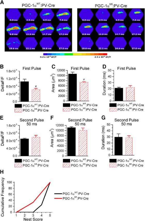
Figure 9.

Deletion of PGC-1α only from parvalbumin interneurons recapitulates circuit dysfunction and behavioral impairment seen in global PGC-1α−/−. A, Example images of the spatiotemporal pattern of voltage-sensitive dye signals evoked in CA1 by Shaffer collateral stimulation in slices from PGC-1αWT:PV-Cre mice and PGC-1αfl/fl:PV-Cre mice on the first pulse. B, The maximum amplitude of the voltage-sensitive dye response is reduced on the first pulse (n = 5, 4). C, The spread of the response is reduced (n = 5, 4). D, The duration of the response is unchanged (n = 5, 4). E, The maximum amplitude of the voltage-sensitive dye response is similar on the second pulse at a 50 ms interval (n = 4, 4). F, The spread of the response is unchanged on the second pulse at a 50 ms interval (n = 4, 4). G, The duration of the response is unchanged on the second pulse at a 50 ms interval (n = 4, 4). H, PGC-1αfl/fl:PV-Cre mice (n = 6) build nests with lower scores compared with PGC-1αWT:PV-Cre mice (n = 8). *p < 0.05, Student's t test.
