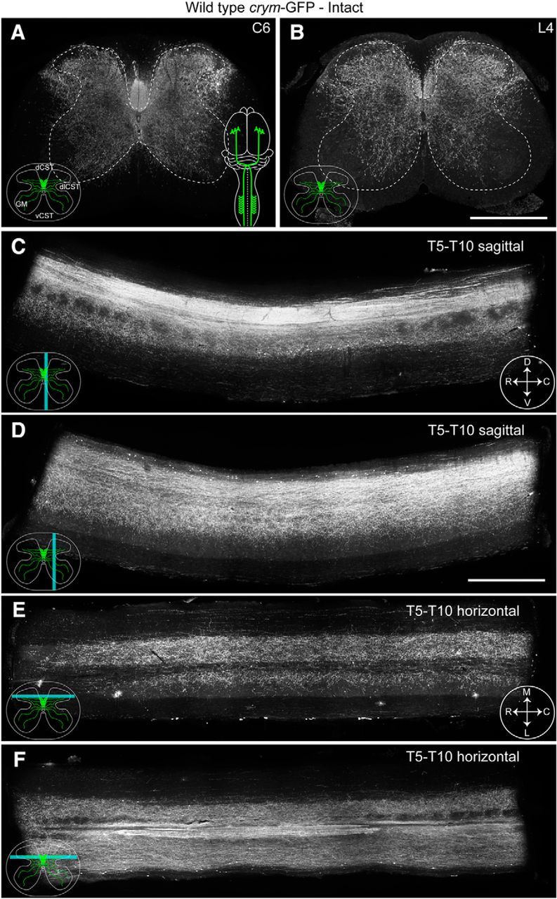
Figure 2.

Intact crym-GFP transgenic mice illustrate comprehensive labeling of the CST. Low-power photomicrographs of transverse sections of cervical (C6, A) and lumbar (L4, B) spinal cord from an intact (color schematic in A shows a bilaterally intact CST in green) adult wild-type crym-GFP mouse show intrinsic GFP expression in the dorsal (dCST), dorsolateral (dlCST), and ventral corticospinal (vCST) tracts and throughout spinal gray matter (GM). Inset schematics (A) reflect intact terminals. Spinal gray and white matter are delineated with a white stippled line (A, B). Sagittal sections through the midline (C, cyan line depicts relative location of section, compass reflects orientation of section: D, dorsal; V, ventral; R, rostral; C, caudal) and mediolateral (D) thoracic cord show intense GFP labeling in the main dorsal CST and GFP+ terminals entering spinal gray matter. Horizontal thoracic sections through the upper (E, cyan line depicts relative location of section, compass reflects orientation of section: M, medial; L, lateral; R, rostral; C, caudal) and lower dorsal horn (F) show GFP+ CST terminals densely localized throughout gray matter. Scale bars: B, 500 μm; D, 1 mm.
