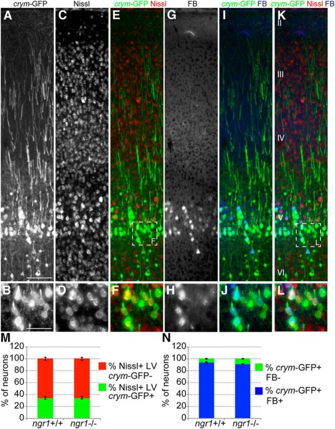
Figure 4.

Crym-GFP is expressed in cortical layer V projection neurons. Photomicrographs (A, B) show robust GFP expression in layer V (cortical layers shown in K) cortical neuron somata, axons and primary dendrites (B, D, F are high-power insets from box shown in E). GFP+ somata are exclusively neuronal as 100% of GFP+ cells are Nissl+ (C–F). Of all Nissl+ cells in layer V (LV), 34.44 ± 2.4 (average number of Nissl+GFP+ somata ± SEM) were GFP+ in ngr1+/+ and 34.93 ± 2.4 were GFP+ in ngr1−/− mice (M). There was no significant difference between genotypes (Student's t test). Unilateral injection of the retrograde tracer FB into the cervical spinal cord resulted in dense labeling of layer V pyramidal neuron somata (G–L) in contralateral cortex 2 weeks after injection (H, J, L are high-power insets from box shown in K). Of all FB+ neurons, 94.69 ± 0.6 were GFP+ in ngr1+/+ and 94.23 ± 0.4 were GFP+ in ngr1−/− mice (N). There was no significant difference between genotypes (Student's t test). Scale bars: A, 100 μm; B, 50 μm.
