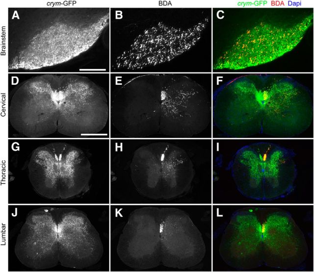
Figure 5.
Intrinsic GFP labeling of the CST in crym-GFP transgenic mice is superior to extrinsic labeling with anterogradely transported BDA. Low-power photomicrographs of brainstem (A–C), cervical (C8, D–F), thoracic (T8, G–I), and lumbar (L4, J–L) spinal cord show robust intrinsic GFP+ CST axons in the medullary pyramid (A) dorsal, dorsolateral and ventral spinal white and gray matter (D, G, J). In contrast, fewer CST axons and terminals are BDA+ in the pyramids (B) and spinal cords (E, H, K) compared with GFP+-labeled axons and terminals in crym-GFP mice (see overlays C, F, I, L; GFP, green; BDA, red; DAPI, blue). Scale bars in A and D, 500 μm.

