Figure 7.

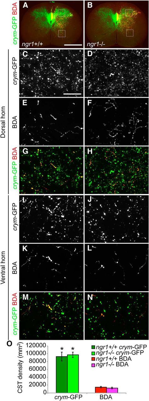
Intrinsic GFP labeling of gray matter CST terminals in crym-GFP transgenic mice is superior to extrinsic labeling with anterogradely transported BDA. Low-power photomicrographs of transverse sections of intact C8 spinal cord from adult ngr1+/+ (A) and ngr1−/− (B) mice show dense GFP+ CST fibers terminating throughout gray matter (A, B, green). BDA+ fibers can be seen exiting the dorsal columns and innervating a more sparse area of gray matter (A, B, red). High-power photomicrographs of the dorsal (C–H) and ventral (I–N) horns show significantly denser GFP+ CST axon terminals (C, D, I, J) compared with BDA+ CST axon terminals (E, F, K, L). Multichannel overlays (G, H, M, N; GFP, green; BDA, red) illustrate the superior efficiency of crym-GFP compared with anterograde BDA tracing. Densitometric analysis showed that significantly more GFP+ CST fibers terminated in intact cervical spinal gray matter compared with BDA+ CST fibers (O, *F(3, 96) = 231.293, p < 0.0005, one-way ANOVA with Bonferonni post hoc comparisons, data are shown as the average density of CST occupation per square millimeter for each label ± SEM). There was no significant difference in the density of crym-GFP-labeled terminals or BDA+ terminals between genotypes. Scale bars: A, 500 μm; C, 10 μm.
