Figure 1.
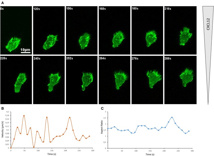
Actin cytoskeleton underlies T cell morphological changes during directional migration. (A) Snapshots of a movie showing a primary CD8+ human T cell expressing Dendra2-LifeAct moving along a CXCL12 gradient created in a collagen IV-coated Ibidi μ-Slide Chemotaxis2D. The cell extends dynamic protrusions and moves toward the source of CXCL12 (top). See Movie S1 in Supplementary Material. The organization of the actin cytoskeleton in T lymphocytes can also be appreciated in previous reports on the dynamics of actin during scanning of target cells (40) and polarization in response to CXCL12 (41). (B) Velocity of the cell shown in (A), calculated for successive 12 s intervals on the basis of the tracking of the cell. (C) Morphology of the cell shown in (A), calculated as Aspect Ratio (length/width of a fitted ellipsis) for each frame.

