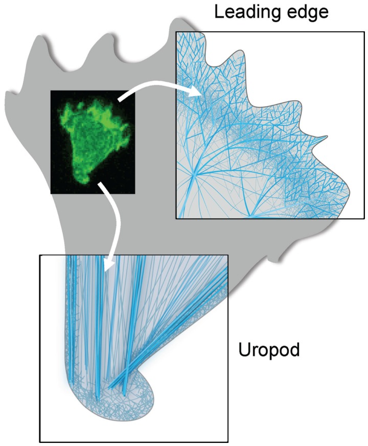
Figure 2.
Actin cytoskeleton organization at the two poles of a migrating T cell. Schematic representations of the ultrastructure of the actin cytoskeleton networks at the leading and trailing edges of the migrating T cell shown in Figure 1. At the leading edge, the T cell that migrates on a 2D surface emits a protrusion that alternates between a lamellipodium and a pseudopodium. It contains a very dynamical and highly branched actin meshwork. At the trailing edge, the T cell uropod is made of a network of parallel actin bundles that can slide along each other to generate contractile forces.

