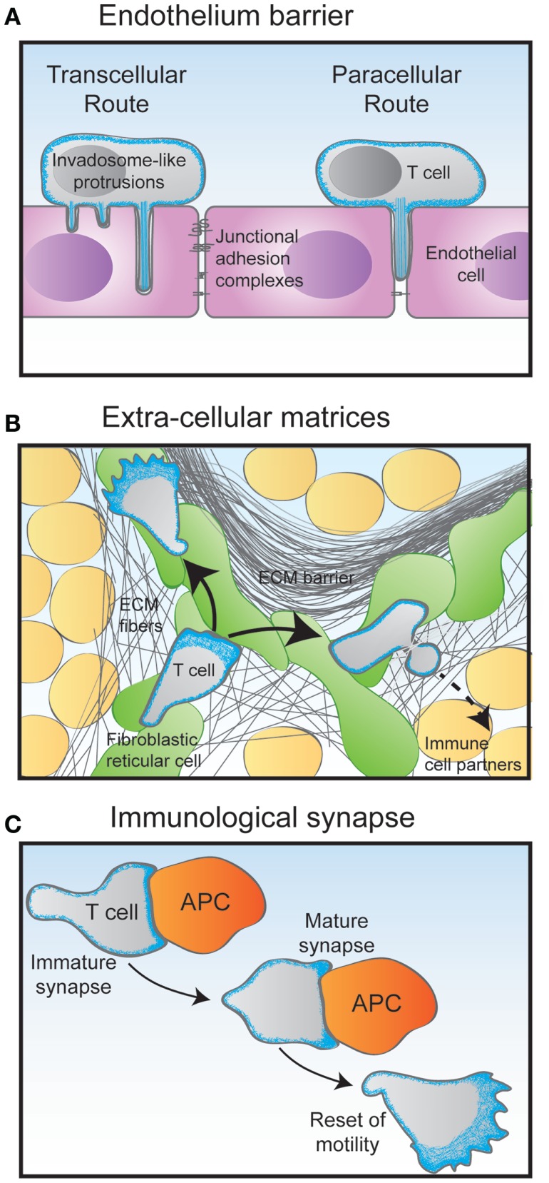
Figure 3.
Migratory challenges faced by T cells during their journey through the organism. (A) The crossing of the endothelium barrier follows steps of T cell tethering and rolling on the luminal surface of the endothelial cells. The combination of chemokines and adhesion molecules triggers firm adhesion to the endothelial surface via the emission of filopodia-like protrusions. Depending on their activation state, T cells then either use the transcellular route by emitting invadopodia-like protrusions that go across the entire endothelial cell body, or the paracellular by squeezing through the junction between two adjacent endothelial cells. (B) Following the crossing of the endothelial barrier and underlying basal membrane, T cells undergo interstitial migration through the tissue they have entered in. Using an amoeboid mode of motility, they crawl and squeeze along and through extracellular matrix (ECM) fibers of various nature. In the lymph node cortex, T cells preferentially migrate along a network of fibroblastic reticular cells decorated with chemokines. (C) During the scanning of antigen-presenting cells (APC), T cells make multiple encounters of various duration and quality. Some contacts may last only a few minutes in the form of an immature immunological synapse. Upon recognition of an APC bearing specific antigens, the T cell stops migrating to assemble a long-lasting immunological synapse. In addition to controlling the interaction between the T cell and the APC, the dynamical actin cytoskeleton serves as a physical platform for numerous signaling events that take place at the immunological synapse to activate the T cell. After a few hours, the activated T cell detaches from its APC partner and regains its motility behavior.

