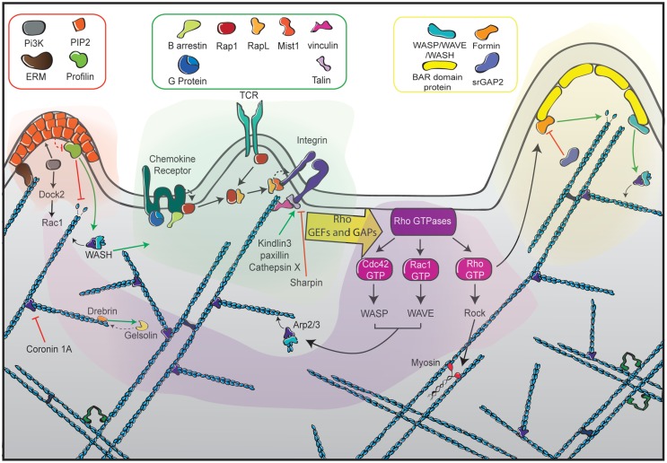
Figure 5.
The different facets of actin cytoskeleton remodeling in migrating T cells. Represented at the center of the scheme (green zone) are the dominant receptors in the control of T cell motility. They include chemokine receptors, the TCR and integrins such as LFA-1, each being interconnected with the actin cytoskeleton with specific sets of signaling molecules. Ligand-mediated triggering of these receptors leads to the activation of the RhoGTPases Rac, Rho, and Cdc42 via GEFs and GAPs (purple zone). Such activation is highly controlled in time and space to orchestrate the assembly of distinct actin networks. Rac activates the WAVE complex, leading to Arp2/3-mediated actin polymerization at the leading edge to form a branched actin network. Cdc42 also favors membrane extension by activating the Arp2/3 complex via WASP. In addition to its major role at the uropod, Rho plays a dual role at the leading edge by promoting actin filament elongation via the formin mDia and by favoring membrane retraction via myosin activation. The left side of the scheme depicts the role of ERM proteins as anchors of the actin cytoskeleton in the plasma membrane (orange zone). The right side of the scheme illustrates the role of BAR-domain proteins as molecular links to guarantee local coordination of membrane curvature and actin polymerization (yellow zone). The actin meshwork is represented as blue filaments that are intertwined with the different signaling areas.

