Figure 5.
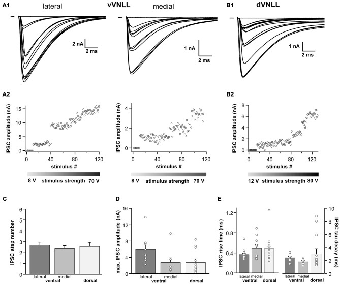
Inhibitory inputs are similar in vVNLL and dVNLL neurons. (A1) Representative IPSC traces of ventrolateral (left) and ventromedial (right) VNLL neurons. Responses were evoked by increasing stimulation strength. Multiple inputs were found both in ventrolateral and ventromedial neurons. All traces represent the average of at least three consecutive recordings to the same stimulus strength. (A2) IPSC amplitude as a function of stimulus number recorded from the ventrolateral and ventromedial vVNLL neurons shown in (A1). (B1) Representative IPSC traces of dVNLL neurons. Responses were evoked by increasing stimulation strength. This neuron shows four distinct current steps. All traces represent the average of at least three consecutive recordings to the same stimulus strength. (B2) IPSC amplitude as a function of stimulus number recorded from the dVNLL neuron shown in (B1). Multiple current steps can be seen. (C) Number of IPSC steps evoked by increasing stimulation strength in vVNLL and dVNLL neurons. VNLL neurons receive two to three inputs. vVNLL: lateral n = 10, medial n = 8; dVNLL n = 12. (D) Maximal IPSC amplitude is similar in all VNLL regions. vVNLL: lateral n = 10, medial n = 8; dVNLL n = 12. (E) Rise time and tau decay time were measured in vVNLL and dVNLL neurons applying a minimal stimulation paradigm. vVNLL: lateral: n = 16; medial: n = 10; dVNLL: n = 14. Data are obtained from 16 C57/Bl6J mice.

