Figure 6.
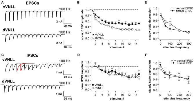
Synaptic short-term plasticity of excitatory and inhibitory inputs is similar in vVNLL and dVNLL neurons. Representative EPSC (A) and IPSC (C) traces in response to 15 stimulations at 100 Hz illustrating short-term plasticity in ventrolateral and dorsal VNLL neurons. Net amplitudes at high frequencies were calculated by fitting a decay function (red trace in C) to the preceding current pulse which was used to set as new baseline. The amplitude of the respective pulse was measured as a subtraction of the maximum and the new baseline (illustrated as red arrow in C). Normalized EPSC (B) and IPSC (D) amplitudes for 100 Hz stimulus in ventrolateral and dorsal VNLL neurons. Steady state depression of EPSCs (E) and IPSCs (F) in ventrolateral and dorsal VNLL neurons. Statistical significance was determined by an unpaired student’s t-test; *p < 0.05. IPSCs: dorsal: n = 9, ventral: n = 9; data are obtained from seven C57/Bl6J mice. EPSCs: dorsal: n = 12, ventral: n = 11; data are obtained from ten C57/Bl6J mice.

