Figure 6.

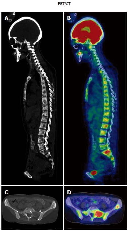
A case of metachronous disseminated carcinomatosis of the bone marrow associated with gastric cancer in a 47-year-old woman: Laboratory findings and positron emission tomography/computed tomography imaging. History: The patient underwent gastric cancer surgery at 36 years of age (histological diagnosis, poorly differentiated adenocarcinoma < signet-ring cell carcinoma), followed by chemotherapy with oral 5-FU (postoperative adjuvant therapy) for 3 years. Eleven years postoperatively, she visited a local orthopedist with a complaint of low back pain. Elevated serum ALP and multiple bone metastases were found on bone scintigraphy, and she was referred to the Shikoku Cancer Center. Laboratory findings: Marked elevation of serum ALP (11740 IU/L) and mild elevation of serum LDH (435 IU/L) were observed with hematological disorders (i.e., DIC and anemia: Hb 6.9 g/dL). Tumor markers (CEA 241 ng/mL; CA19-9 212 U/mL) and bone metabolic markers (1CTP 17.7 ng/mL; Urine NTx > 300 nmol BCE/L; BAP 1260 μg/L) were elevated. These findings were suggestive of recurrence of the gastric cancer in the bone (i.e., disseminated carcinomatosis of the bone marrow). PET/CT imaging: Osteolytic changes with FDG accumulation were observed in most vertebrae. A, B: Sagittal views of CT (A) and PET/CT fusion images (B); C, D: Transaxial views of the sacrum (S1) on CT (C; arrowheads, osteolytic change) and PET/CT fusion image (D). PET: Positron emission tomography; CT: Computed tomography.
