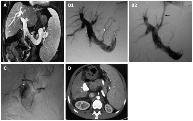
Figure 1.

Patient with hepatitis C cirrhosis associated with esophageal gastric-fundus variceal bleeding was diagnosed with hepatocellular carcinoma 37 mo after transjugular intrahepatic portosystemic shunt. We treated HCC with TACE. A: Obvious portal vein dilation (white arrow) revealed by magnetic resonance cholangiopancreatography; B1: Obvious portal vein dilation (short white arrow) and gastric coronary vein varicosis (long white arrow); B2: Gastric coronary vein embolism (long white arrow) and distributary channel (short white arrow); C: Tumor lesion with rich blood supply in the right hepatic lobe 37 mo after TIPS by hepatic angiography (white arrow); D: Iodine oil deposited in the lesion after TACE, shown by contrast-enhanced CT (white arrow). HCC: Hepatocellular carcinoma; TACE: Transarterial chemoembolization; TIPS: Transjugular intrahepatic portosystemic shunt; CT: Computed tomography.
