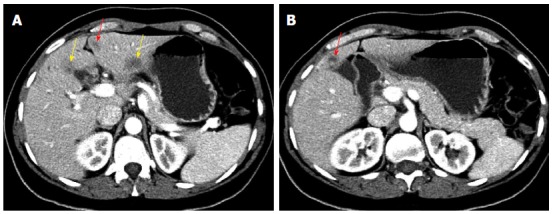
Figure 2.

Computed tomography of abdomen. A: Axial contrast enhanced (portal phase, liver window, 200/50) demonstrates three hypodense lesions. The previously seen segment III and IVB lesions have increased in size (yellow arrows) and the one medial to the falciform ligament is now apparent (red arrow); B: Axial contrast enhanced (portal phase, liver window, 200/50) demonstrates a new liver lesion (arrow).
