
Figure 3.
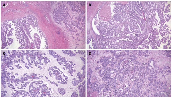
Microscopic features of the tumor. A: Intraductal papillary mass with adjacent invasive adenocarcinoma; B: The papillary mass with fine vascular cores was lined by foveolar type epithelium; C: Some areas showed mucin production; D: The metastatic masses in the liver showed infiltrative features.