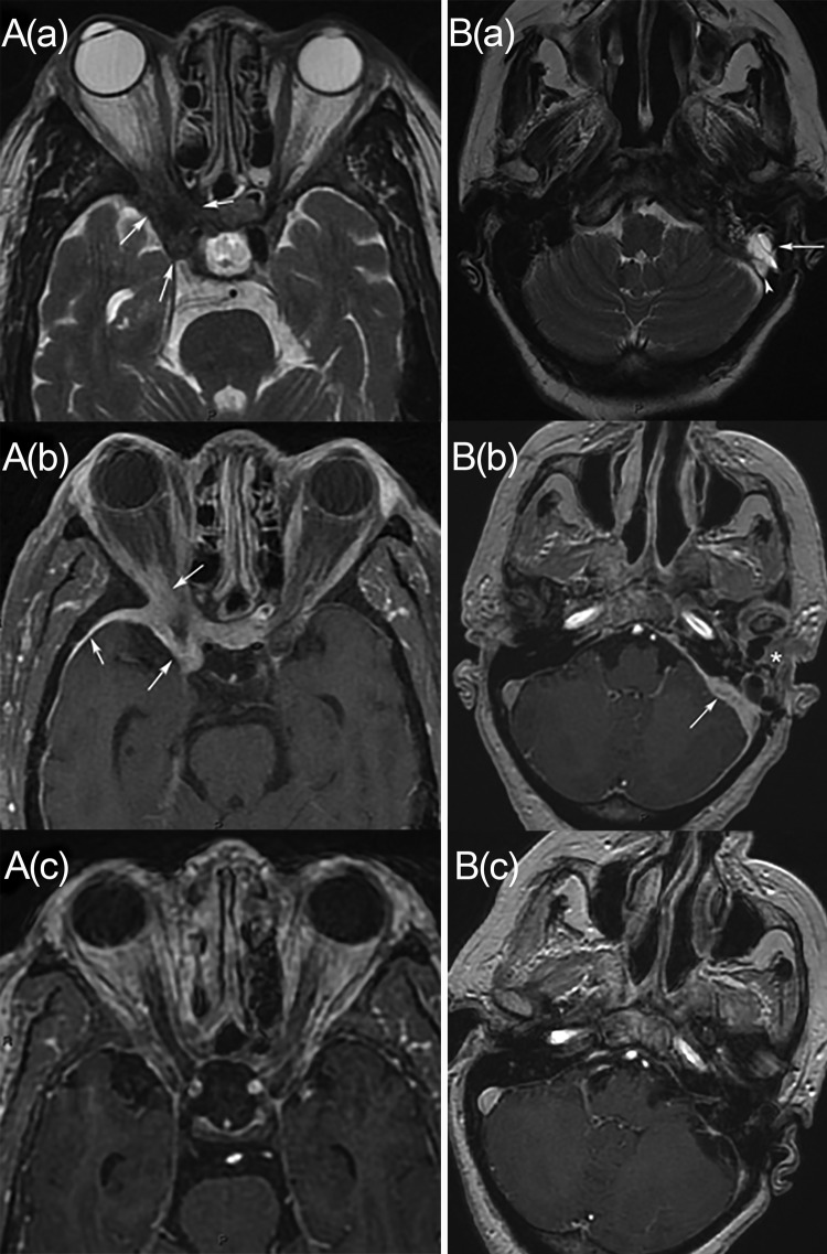
FIG 1.
Two cases of meningeal invasive aspergillosis. (A) Case 1, involving a 71-year-old woman. (a and b) Axial T2-weighted (a) and axial enhanced T1-weighted (b) MRI images show soft tissue thickening in the region of the right cavernous sinus and orbital apex (arrows). There is also meningeal enhancement adjacent to the thickening (arrowhead). (c) An axial enhanced T1-weighted MRI study performed after treatment shows important decreases in soft tissue thickening and meningitis. (B) Case 2, involving a 68-year-old woman. (a) An axial T2-weighted MRI image shows left filling of the mastoid air cells (arrow) and dural venous sinus thrombosis (arrowhead). (b) An axial enhanced T1-weighted MRI image shows soft tissue thickening involving the left temporal bone and the mandibular region (asterisk), with meningitis (arrow). (c) A follow-up axial enhanced T1-weighted MRI image shows an important decrease in the involvement and persistent dural venous sinus thrombosis.

