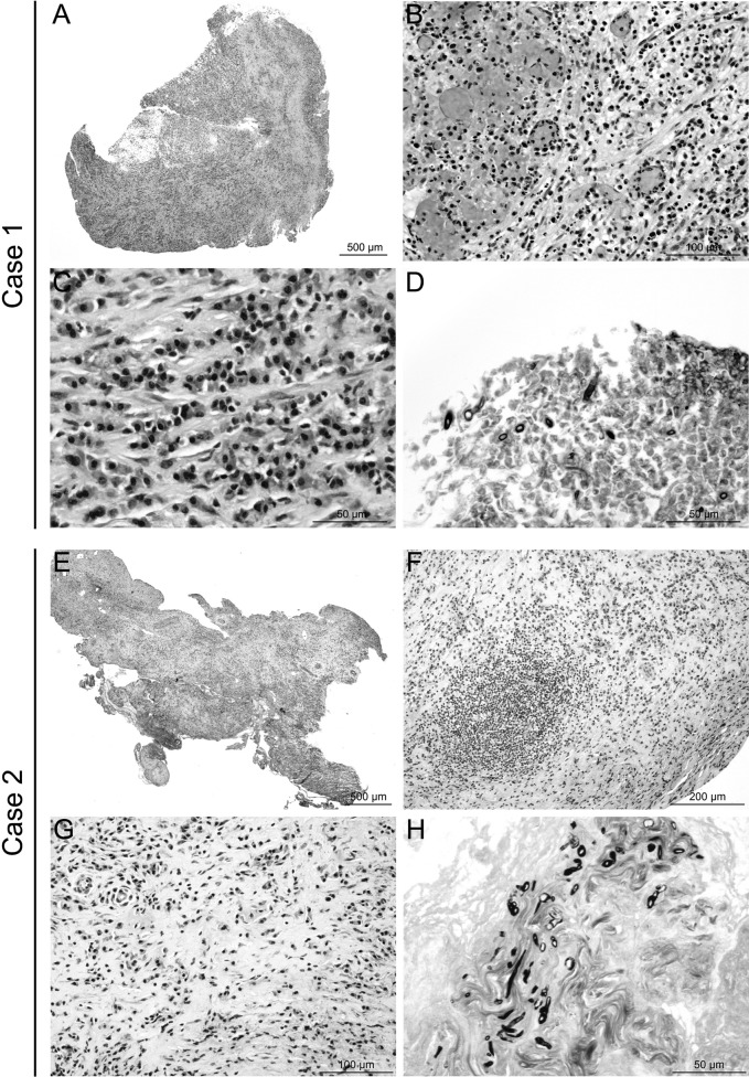
FIG 2.
Histological lesion profiles and fungus identification. (A to D) Case 1, retroorbital biopsy samples. A diffuse subacute inflammatory lesion was observed (A), characterized by infiltration of neutrophils, macrophages (B), lymphocytes, and plasma cells (C), sometimes associated with vascular and ischemic necrosis, with hemorrhage (B). Rare fragmented hyphae were detected (size, <50 μm) (D). These hyaline hyphae were septate but no branching was identified, probably because of the small sizes of the samples. (E to H) Case 2, meningeal biopsy samples. A diffuse chronic inflammatory lesion was observed (E), characterized by infiltration of macrophages, lymphocytes, and plasma cells, sometimes organized in pseudolymphoid follicles (F), included in dense mature connective tissue (G). Small colonies of irregularly dispersed hyaline hyphae were identified (H). These hyphae were septate and branching, with the branches forming acute angles. Both lesion presentations were consistent with aspergillosis. Hematoxylin-eosin staining (A, B, C, E, F, and G) or Gomori-Grocott staining (D and H).

