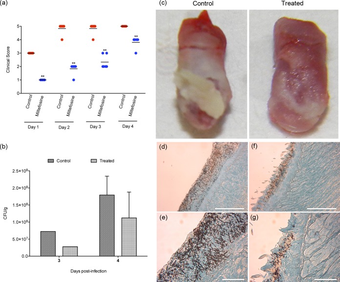
FIG 2.
In vivo evaluation of the protective effect of miltefosine against oral candidiasis. Treated groups received 50 mg/kg of miltefosine, twice a day, by topical administration. Miltefosine treatment was initiated the day before infection (day −1). The control group received saline twice a day, also by topical administration. (a) Clinical score of candidiasis progression, showing that miltefosine has a protective role, preventing the development of disease. **, P < 0.01; statistical analysis by nonparametric Mann-Whitney test. (b) Extent of C. albicans colonization on tongues from control mice and mice treated with miltefosine. Only one animal from each group was sacrificed at the end of day 3. Bars indicate the standard deviations. The graph shows the reduction in C. albicans colonizing the tongues of animals treated with miltefosine in comparison to the untreated control group. Statistical analysis by nonparametric Mann-Whitney test (4th day), P = 0.0952, and Mann-Whitney U test, 4.000. (c) Visual analysis of the extension of tongue colonization by C. albicans in mice from the control group (left) and the group treated with miltefosine (right) at the end of day 4. Control tongues showed a dense biofilm covering the entire tongue surface on day 4 (left), and miltefosine treatment showed a protective role by reducing the tongue colonization and reducing the biofilm formation (right). (d to g) Histological sections of tongues from control mice (d and e) and mice treated with miltefosine (f and g) sacrificed on the 4th day and stained with Grocott-Gomori stain and silver methanamine. The administration of miltefosine reduced tissue colonization, inhibited hypha formation, and reduced invasive behavior at the infection site (f and g).

