
FIG 2.
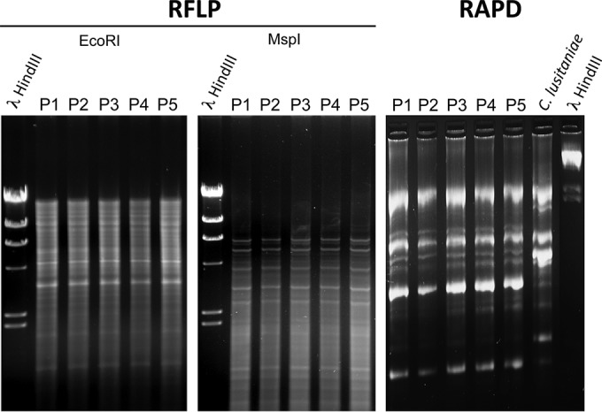
RFLP and RAPD analysis of C. lusitaniae isolates P1 to P5. RFLP analysis was carried out with EcoRI and MspI. RFLP profiles are shown in Fig. S1 in the supplemental material. RAPD analysis was performed as described in Materials and Methods. Identical patterns of ethidium bromide-stained profiles suggest a high-level relationship between the strains. Lambda phage DNAs digested by HindIII were loaded as standard sizes. A separate C. lusitaniae isolate (Sanglard laboratory collection) was used as a control for RAPD analysis.