FIG 3.
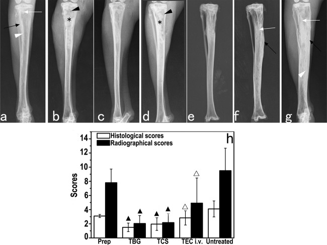
(a to g) Representative radiographs of rabbit tibiae before and after treatment. (a) Experimental MRSA-induced tibia osteomyelitis shows bone destruction (white arrows), periosteal new bone formation (black arrows), and sequestral bone formation (white arrowheads). (b and d) Bone windows (black arrowheads) and implantation of TBG (b) or TCS (d) implants (stars) can be observed 1 day after debridement. (c and e to g) Healing of osteomyelitis was observed 6 weeks after implantation of TBG (c) or TCS (e) implants, but more progression was observed for the intravenous teicoplanin (f) and untreated (g) groups. (h) Radiographic and histological osteomyelitis scores for rabbit tibiae. ▲ indicates a significant difference compared with other groups, and △ indicates a significant difference compared with the untreated group (P < 0.05).

