FIG 4.
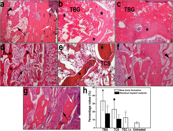
Representative images of H&E-stained sections of rabbit tibiae before and after treatment. (a) Typical signs of bone infection were observed after induction of osteomyelitis, including the destruction of bone (white arrow), fibrosis (black arrow), periosteal new bone formation (black arrowhead), and intramedullary abscess (white arrowhead) (magnification, ×40). (b and c) Tibiae treated with the TBG implants show newly formed bone (stars) located intimately around and within the degraded TBG implant, with no signs of chronic inflammation (magnifications, ×40 and ×200 [c]). (d and e) Tibiae treated with the TCS implants show degraded CaSO4 surrounded by a moderate amount of new bone (stars), accompanied by obvious chronic fibrosis with proliferated foamy histiocytes and multinucleated granulocytes (black arrow) (magnifications, ×40 [d] and ×200 [e]). (f and g) Moderate to severe inflammation was observed in rabbits treated with teicoplanin intravenously (magnification, ×40) (f) and in the untreated group (magnification, ×40) (g), such as destruction of bone (white arrows), fibrosis (black arrows), and periosteal new bone formation (black arrowhead). (h) Quantitation of new bone and residual amounts of the implant from H&E-stained sections. ▲ indicates a significant difference compared with other groups, △ indicates a significant difference compared with the TCS group, ■ indicates a significant difference compared with the intravenous treatment and untreated groups, and □ indicates a significant difference compared with the untreated group (P < 0.05).

