
FIG 4.
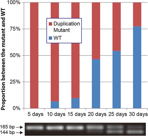
Reversion of a duplication mutation. The proportional variation between the wild type and a strain with a duplication mutation (M-Dup1) during the course of a 30-day incubation without antibiotic pressure is shown in a bar graph. Changes in PCR products containing the duplication region are shown below the graph.