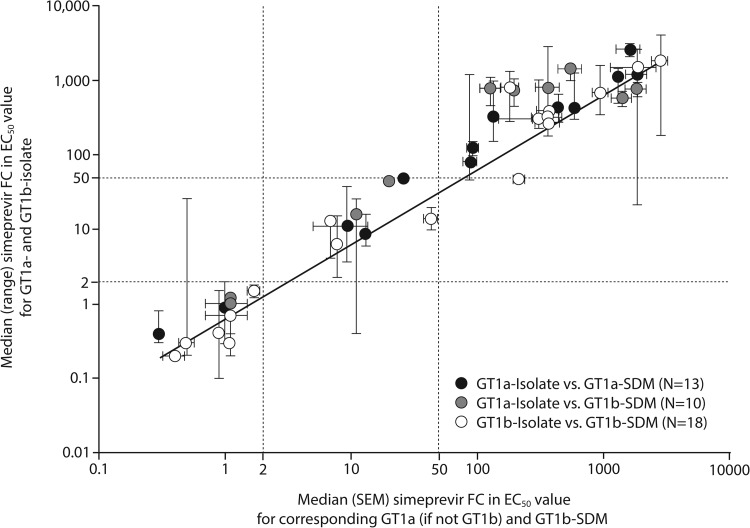
FIG 3.
Correlation of the median simeprevir FC values for HCV genotype 1a and genotype 1b isolates carrying amino acid substitutions at NS3 positions 80, 122, 155, 156, 168, and/or I/V170T, with the median simeprevir FC values for the corresponding SDMs in a genotype 1a or genotype 1b replicon backbone. Black/gray and white symbols, genotype 1a and genotype 1b isolates/SDMs, respectively. Simeprevir FC values for individual clinical isolates were calculated as median values across 1 to 7 replicates (the majority [341 of 435] of isolates were tested ≥3 times). Clinical isolates containing the same single, double, triple, or no amino acid substitutions at NS3 positions 43, 80, 122, 155, 156, 168, and/or 170 were grouped, and the median FC value for each group was determined (each group contained 1 to 95 individual isolates; for details, see Table S5 in the supplemental material). Simeprevir FC values for SDMs carrying corresponding simeprevir resistance-associated amino acid substitutions were calculated as median values across 1 to 24 experimental replicates (the majority [38 of 41] of SDMs were tested ≥3 times). EC50, 50% effective concentration; GT, genotype; FC, fold change; HCV, hepatitis C virus; SDM-1a or -1b, site-directed mutant assessed in an HCV genotype 1a or genotype 1b replicon backbone, respectively; SEM, standard error of the mean.

