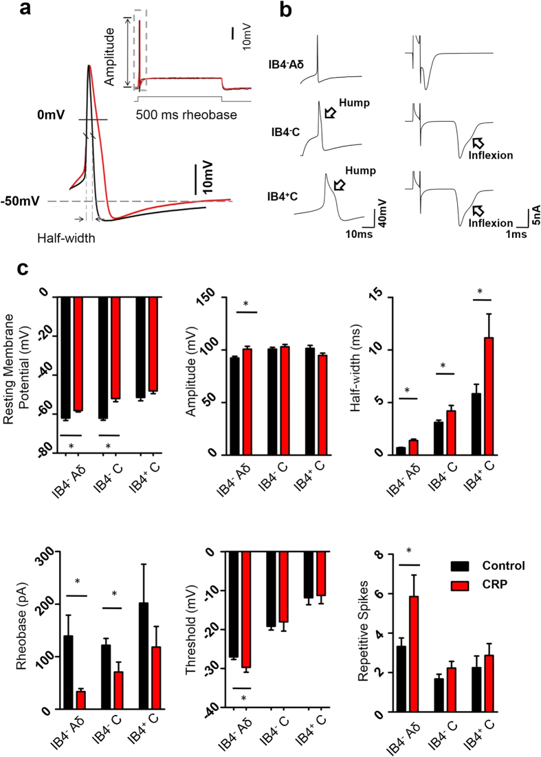
Figure 3. Membrane property and cell excitability of different subsets of nociceptive DRG neurons in control and CRP rats.
(a) A representative action potential of whole-cell configuration evoked by a depolarizing current injection from control (in black) and from CRP (in red) group. (b) Classification of all small DRG neurons recorded. Note that three subtypes were determined by IB4 staining, shape of action potential (left panels) and type of the afferent. A single electrical stimulation of dorsal root 3 mm away from DRG was applied to distinguish C type afferent from Aδ type. (c) Showing resting membrane potential (RMP), action potential (AP) properties, such as amplitude, half-width, rheobase and threshold as well as repetitive firing in response to current injection in IB4− Aδ-, IB4− C- and IB4+ C-type DRG neurons from control and CRP rats. All data are expressed as mean ± S.E.M. *P < 0.05 indicates statistically significant differences between control and CRP rats (ANOVA).

