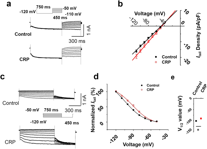
Figure 7. CRP changed voltage dependence of Ih activation in IB4− Aδ type neuron, but not the reversal potential.
(a) Reversal potential of Ih from control (middle panel) and CRP IB4− Aδ type neurons (lower panel) were achieved by first applying a prepulse to −120 mV to fully activate Ih and then examining the tail currents after repolarization to test potentials from −110 to −50 mV (upper panel). Tail currents were plotted against test potentials. (b) No difference of reversal potential of Ih current was shown between control (black) and CRP (red) group. (c) The activation curve of Ih from control (upper panel) and CRP IB4− Aδ neurons (lower panel) was constructed by measuring tail currents at −120 mV after application of prepulse potentials between −50 to −120 mV (middle inset) and fitted with a Boltzmann equation. (d) The midpoint (V1/2) for activation of Ih in the CRP IB4− Aδ neurons (red) was shifted 11 mV in the depolarizing direction compared to control neurons (black). (e) The difference of V1/2 for activation of Ih between two groups was significant. All data are expressed as mean ± S.E.M. *P < 0.05 as compared to control rats.

