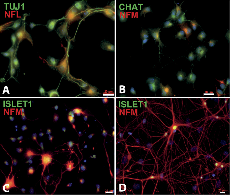
Figure 1. Characterization of purified MNs obtained from E14.5 embryo spinal cord of CD-1 mice.
MNs were extracted from E14.5 embryos and characterized by immunofluorescence staining using the TUJ1 (A) in green), NFL ((A), in red) and NFM ((B–D) in red) neuronal markers and the CHAT (B) in green) and Islet1 ((C,D) in green) specific MN markers after 2 days of culture on a poly-D-lysine coated cover glass (200,000 cells/cm2). NFM (in red) and Islet1 (in green) positive cells with long neurites can be observed after cultivating MNs for 14 days (D). Scale bars 20 μm.

