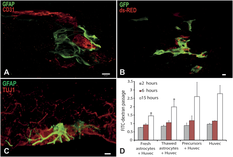
Figure 4. Characterisation of astrocyte functionality obtained from adult spinal cord of 5-month-old wtSOD1mice.
Astrocytes were cocultured with endothelial cells and fibroblasts in a three-dimensional culture system in which capillary-like tubes form (A,B). Astrocytes were characterized by GFAP expression in green (A,C), or transduced to express ds-Red (B). Endothelial cells were detected by CD31 expression in red (A) or transduced to express GFP in green (B). Astrocytes were cocultured with MNs in a three-dimensional culture system promoting axonal migration. Astrocytes were identified using GFAP expression in green, and MNs using TUJ1 expression in red (C). The capacity of astrocytes to decrease endothelial cell permeability was assessed by quantification of FITC conjugated dextran diffusion through an endothelial cell monolayer, compared to spinal cord-derived oligodendrocyte progenitor cells as a control. Astrocytes significantly decreased FITC-dextran diffusion through endothelial cell monolayer after 15 hours of contact compared to endothelial cells alone or endothelial cells cocultured with oligodendrocyte progenitor cells (p < 0.005; n = 3). Scale bar 10 μm.

