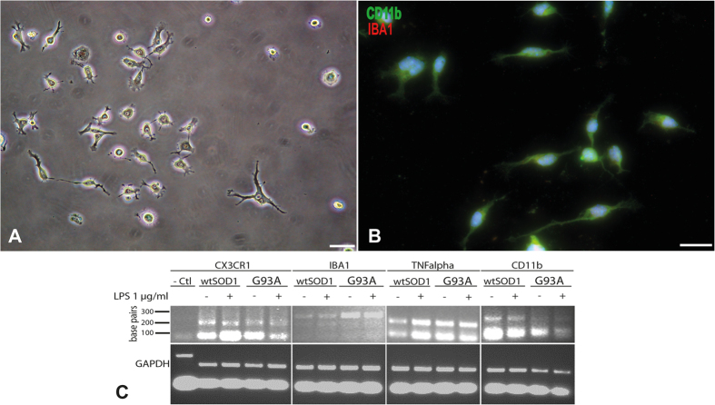
Figure 5. Characterisation of microglia purified from adult spinal cord of 5-month-old mice.
Phase contrast microscopy of purified microglia cells after 14 days of culture (20,000 cells/cm2) (A). Immunocytochemical characterization of purified microglia using CD11b in green and IBA1 in red (B). Scale bar 20 μm. Qualitative PCR showing expression by microglia from wtSOD1 and SOD1G93A mice of specific markers such as CX3CR1, IBA1, TNFalpha and CD11b, after activation or not with LPS (C). Urothelial cells were used as negative control.

