Figure 3.
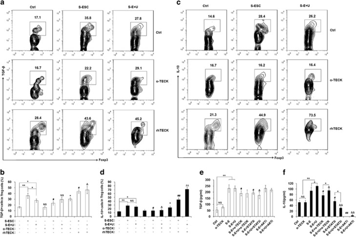
TECK promotes IL-10 and TGF-β in Tregs. After activation with anti-CD3 and anti-CD28 antibodies for 48 h, naïve T cells were cultured with S-ESC or S-E+U in the presence or absence of anti-human TECK neutralizing Abs (α-TECK) (5 μg/ml) or recombinant human TECK (rhTECK) (100 ng/ml) for 5 days, then further incubated with ionomycin (100 ng/ml), PMA (100 ng/ml) and BFA (10 μg/ml) for 4 h. The levels of intracellular TGF-β (a, b) and IL-10 (c, d) in CD4+Foxp3+Tregs were analyzed by flow cytometry. CD4+CD25+ regulatory T cells were isolated from the peripheral blood of healthy fertile women by MACS and incubated with S-ESC, S-E+U, anti-TECK neutralizing Abs, STAT3 (10 μM) and/or AKT inhibitors (30 μM). The amount of TGF-β (e) and IL-10 (f) secreted by the Tregs into the supernatant was determined. Data are expressed as the mean±S.D. *P<0.05 or **P<0.01 compared with medium/vehicle control; #P<0.05, ##P<0.01 compared with S-ESC group without α-TECK and rhTECK; ΔP<0.05, ΔΔP<0.01 compared with the S-E+U group without α-TECK and rhTECK

