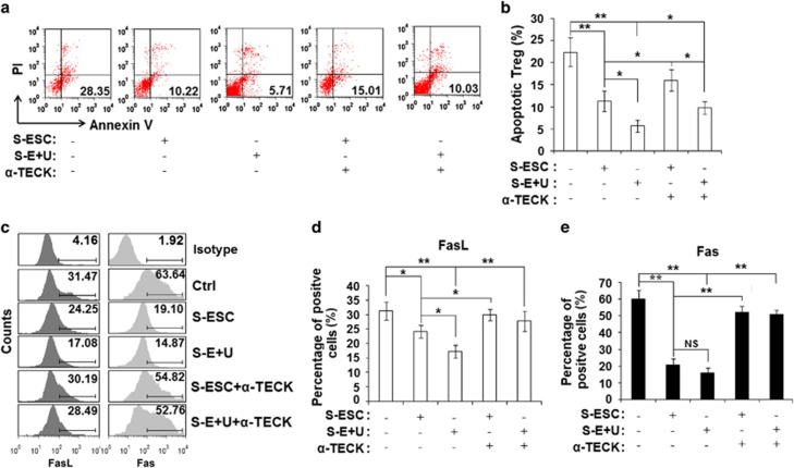
Figure 4.
TECK represses Treg apoptosis. CD4+CD25+ Tregs were isolated from the peripheral blood of healthy women by MACS and cultured in S-ESC or S-E+U with or without α-TECK for 48 h. The Tregs were then stained for the Annexin V-FITC assay (a, b), and the percentages of FasL+ (c, d) and Fas+ Treg cells (c, e) were determined. Data are expressed as the mean±S.D.

