Figure 6.
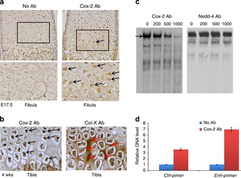
Cox-2 expression and interaction with Col10a1 cis-enhancer. (a) IHC assay using Cox-2 antibody on sagittal sections of mouse limb (fibula) at E17.5 showed that Cox-2 is strongly expressed in nuclei of hypertrophic chondrocytes but not in resting or proliferative chondrocytes (black box and arrows). Bottom showed higher magnification of the boxed area. Left panel shows no antibody control. (b) IHC assay using Cox-2 antibody on tibia sections at 4 weeks' age also detected high-level Cox-2 expression in nuclei of hypertrophic chondrocytes (left panel, black arrows). Right panel shows IHC assay of collagen X, which is mostly expressed extracellular within hypertrophic zone (red arrows). (c) EMSA assay detected specific binding complex (black arrow) formed by the Col10a1 enhancer and hypertrophic MCT cell nuclear extracts as previously described.38 The signal intensity decreased when 200 ng of Cox-2 antibody was added, 500 ng of antibody further reduced the signal, whereas 1000 ng of antibody only showed faint signal (left panel). No signal reduction was seen in parallel experiment in which gradient amount of Nedd4 antibody was used (right panel). Data of non-biotinylated competitor control were not shown. (d) Illustrated is the result of ChIP experiment showing precipitated DNA enriched by Cox-2 antibody or control IgG. qPCR using primers flanking the enhancer suggested a significant enrichment (~7-fold, P=0.034) of the enhancer by Cox-2 antibody, whereas sequence flanking the intronic control region did not show significant enrichment (~3-fold, P=0.062)

