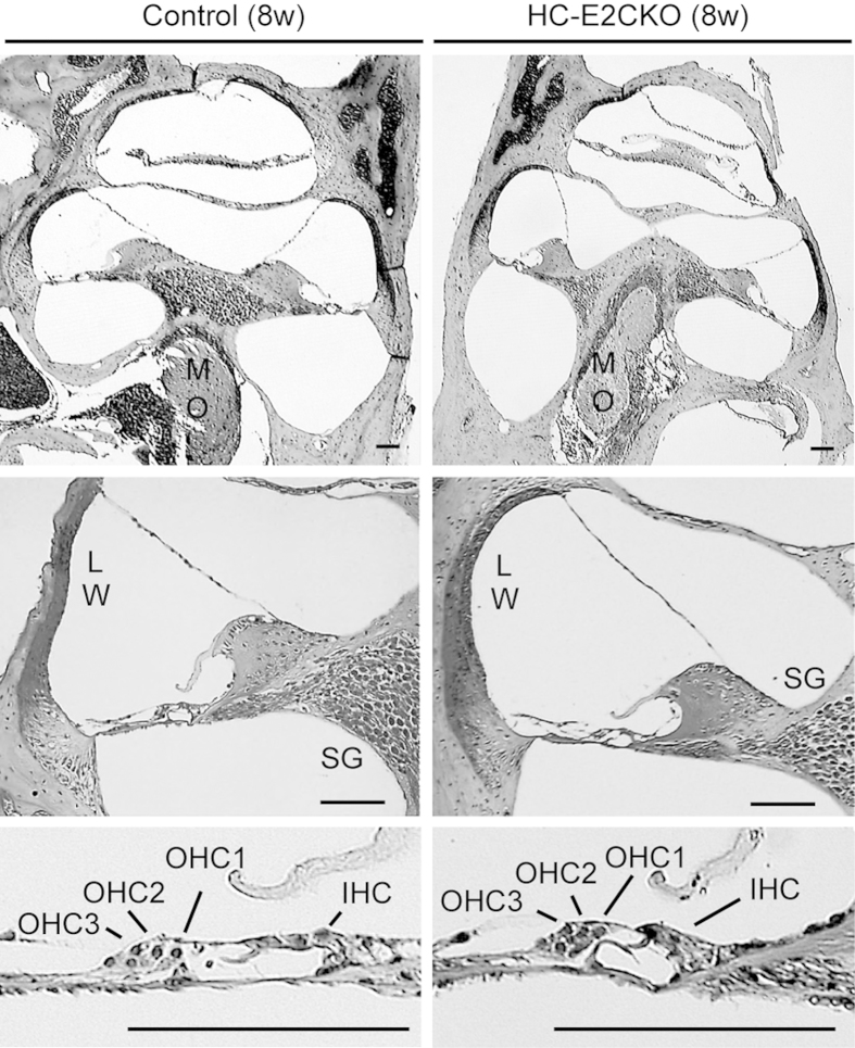
Figure 2. Cochlear morphology was normal in HC-E2CKO mice.
Representative photomicrographs of cochleae stained with hematoxylin and eosin from 8-week-old control and HC-E2CKO mice. No significant difference was detected in the cochleae, including the lateral wall (LW), spiral ganglion (SG) in modiolus (MO), and inner and outer hair cells (IHCs, OHCs). Scale bar, 50 μm.

