Figure 3.
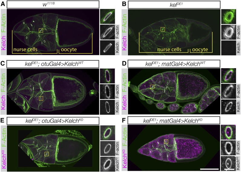
Kelch protein resistant to autocatalytic degradation is fully functional. (A and B) Confocal maximum intensity projections of egg chambers labeled with Kelch antibody and fluorescent phalloidin. (A) Wild-type stage 10 egg chamber. Inset shows higher-resolution image of a ring canal, revealing the F-actin cytoskeleton and associated Kelch protein. (B) Stage 10 egg chamber from a kelDE1 female. The oocyte is considerably smaller than the comparably staged wild-type egg chamber in A, ring canals lack detectable Kelch protein, and the F-actin cytoskeleton largely occludes the lumens of the ring canals. (C and D) Ovaries from kelDE1 rescued with a wild-type cDNA driven at low (otuGal4, C) or high (matGal4, D) levels of expression. kelDE1 egg chambers rescued with otuGal4 resemble wild-type egg chambers. Ovaries rescued with high-level Kelch expression using matGal4 accumulate cytoplasmic aggregates of Kelch (D), but Kelch localizes to ring canals and rescues the cytoskeletal defects. (E and F) KelchK0 rescues the kelch ring canal cytoskeletal phenotype at both low (E, otuGal4) and high (F, matGal4) levels of expression. Accumulation of Kelch cytoplasmic aggregates was greatly enhanced with matGal4-mediated expression (F). Scale bars, 50 µm for egg chamber images and 10 µm for cropped ring canal images.

