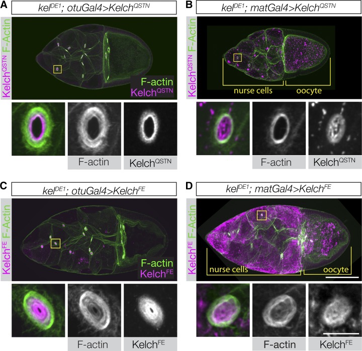
Figure 6.
Disrupting the Kelch–Cul3 interaction results in a failure to rescue the kelch mutant phenotype. (A–D) Confocal projections of egg chambers labeled with Kelch antibody and fluorescent phalloidin. (A and B) Egg chambers expressing KelchQSTN rescue the F-actin cytoskeletal defects of the kelDE1 mutant. (A) Expression at lower levels using otuGal4 occasionally resulted in an incomplete rescue, with some accumulation of KelchQSTN biased toward the ring canal lumen and an F-actin phenotype intermediate between wild type and kelch-like. (B) High-level expression of KelchQSTN driven by matGal4 fully rescued the ring canals with respect to F-actin organization. However, matGal4-driven KelchQSTN formed abundant cytoplasmic aggregates and tended to form aggregates of Kelch in the ring canal lumens (high magnification ring canal images in B). (C and D) Expression of KelchFE at either low (C) or high (D) levels failed to rescue the kelDE1 F-actin cytoskeletal defects (compare high-magnification ring canal images with those in Figure 3B). In addition, the KelchFE mutant protein accumulated in the ring canal lumen, colocalizing with the disorganized F-actin. When driven by matGal4, KelchFE also formed highly abundant aggregates throughout the germline cytoplasm. Scale bars, 50 µm for egg chamber images and 10 µm for cropped ring canal images.

