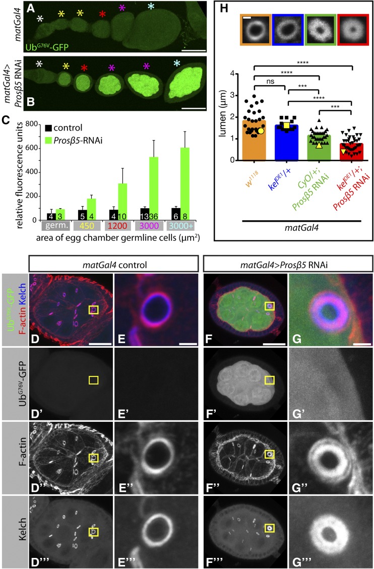
Figure 7.
Proteasome inhibition by RNAi leads to kelch-like ring canals. (A–C) Proteasome inhibition by RNAi is effective as evidenced by UbG76V–GFP reporter protein accumulation. Representative images showing UbG76V–GFP reporter protein levels in control egg chambers (A) or egg chambers expressing shRNAs targeting Prosβ5 proteasome subunit with matGal4 driver (B). (C) Quantification of UbG76V–GFP reporter protein levels. Mean GFP fluorescence of egg chamber germline cell area was measured for individual egg chambers. Egg chambers were grouped based on size (area, μm2) and the average GFP fluorescence for each area grouping is plotted. Area groupings on the x-axis indicate the maximum area cut-off for each particular group (e.g., 3000 μm2 group represents egg chambers 1201–3000 μm2 in size). Color-coded asterisks in A and B designate egg chambers that are representative of the area groupings plotted in C. The sample size is indicated at the base of each bar, and error bars represent standard deviation. (D–G) Representative images of egg chambers (D and F) or ring canals (E and G) expressing UbG76V–GFP reporter protein (D’–G’) and stained for F-actin (D′′–G′′) and Kelch (D′′′–G′′′). Note that the ring canal in G is kelch-like with a thicker F-actin ring (G”). (H) Reduction of one copy of kelch dominantly enhances the kelch-like ring canal phenotype observed upon proteasome inhibition. (Top) Representative images of ring canals stained for F-actin with color-coded boxes matching genotype displayed in graph below. (Graph) Quantification of ring canal lumen span (see Figure S5 for lumen span measurements). Bars represent mean of lumen span and black points represent all individual measurements. Yellow points correspond to individual ring canals shown in top panels. (***) P < 0.0005, (****) P < 0.0001, one-way ANOVA, Tukey’s multiple comparison test. E and G are insets of yellow boxes in D and F, respectively. Scale bars, 50 μm (A and B), 20 μm (D and F), 2 μm (E and G), 1 μm (H).

