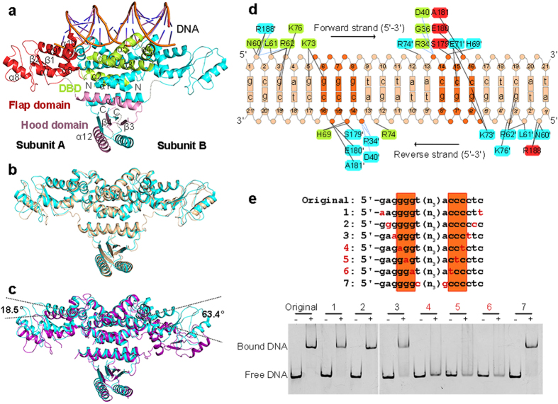
Figure 2. Structure of HetR−DNA complex.
(a) Overall structure of Anabaena HetR dimer in complex with the 21-bp DNA from the hetP promoter. The secondary structural elements of DBD (limon), flap domain (red) and hood domain (pink) in subunit A are labeled. The missing residues are depicted as the dashed lines. Comparison of DNA-bound Anabaena HetR (cyan) with (b) the DNA-bound (wheat) and (c) apo-form (purple) Fischerella HetR. The rotation angles of the flap domains are labeled. (d) Schematic representation of interaction networks between Anabaena HetR and DNA. Interacting residues are marked with the same color as their corresponding domains, respectively. Direct hydrogen bonds are indicated as black lines and water-mediated hydrogen bonds as blue dashes. (e) EMSA assays of HetR with variations of DNA. DNA samples with and without HetR are shown as “+” and “−”, respectively. The corresponding DNA variations numbered as No.1 to 7. The 5′-g6gg and 5′-c14cc motifs are highlighted in dark orange.

