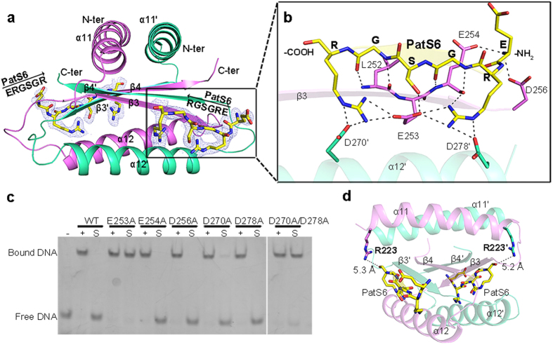
Figure 3. PatS6-binding mode.
(a) Overall structure of the PatS6−HetRHood dimer. Two PatS6 peptides (yellow) are shown as sticks, with the Fo − Fc electron-density omit map contoured at 3.0 sigma. (b) A close-up view of the PatS6-binding site. Hydrogen bonds and the polar interactions are indicated as dashed lines. (c) EMSA assays of HetR and mutants with original DNA sequence, in the presence or absence of PatS6. “−” represents DNA probe without protein. “+” indicates DNA incubated with HetR or mutants, whereas “S” indicates addition of PatS6 to the HetR−DNA complex. (d) The distance between Arg223 and the C-terminus of PatS6 in each subunit is labeled.

