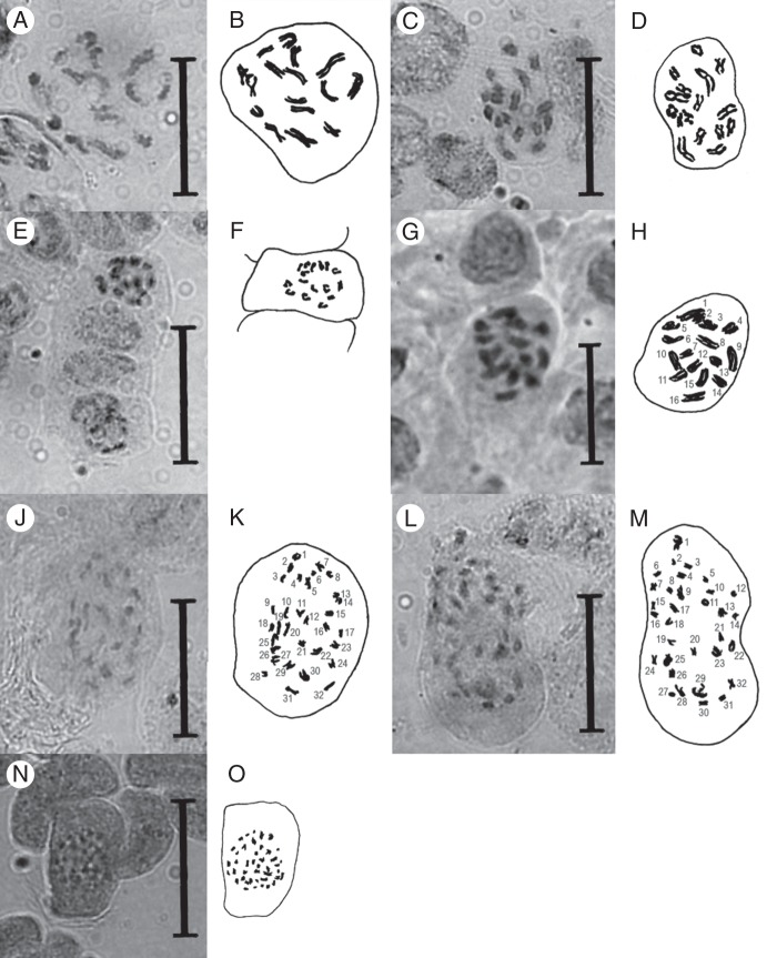
Fig. 2.
Light microscope photographs and camera lucida drawings of orcein-stained mitotic prophase plates from rhizophyll tip meristems of Genlisea spp. (A, B) G. uncinata, 2n = 16. (C, D) G. metallica, 2n = 16. (E, F) G. violacea from Couto de Magalhães, 2n = 16. (G, H) G. flexuosa, 2n = 16. (J, K) G. hispidula, 2n = 32. (L, M) G. subglabra, 2n = 32. (N, O) G. guianensis, 2n ∼40. Scale bars = 3 μm.

