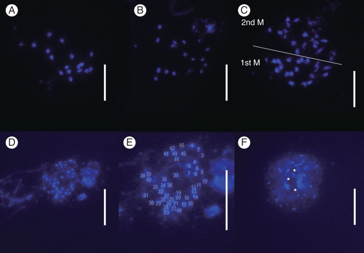
Fig. 3.
Metaphase chromosomes from pollen mother cells of Genlisea spp., stained with DAPI. (A–C) G. margaretae from Zambia, n = 18, 19. (C) Two meiotic metaphase cells. (D–F) G. aurea var. minor from Itacambira, pre-meiotic mitosis cells, 2n = 46. Note the smaller chromosome size compared with G. margaretae. (F) DAPI-stained interphase nuclei. Nucleoli (*) can be identified as DAPI-negative regions surrounded by more DAPI-positive DNA staining. Scale bars = 10 μm.

