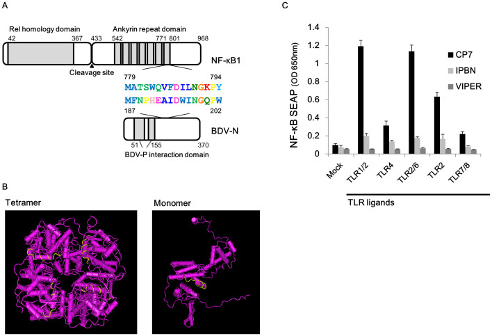
Figure 2. Inhibitory effect of the virus peptide on NF-κB activation.
(A) Schematic diagram of NF-κB1 and BDV-N. (B) The crystallographic structure of the BDV-N tetramer and monomer (Protein Databank # 1N93). Yellow lines indicate the IPBN. (C) Inhibitory effect of the viral peptide on NF-κB activation. THP1-CD14 cells were pre-treated with 100 μg/ml of the viral peptide derived from BDV-N, a negative control peptide (CP7), or the positive control peptide (VIPER) and stimulated with five TLR ligands. At 24 h post-stimulation, the SEAP activities in the supernatants were measured. Error bars represent the standard deviation of the mean (N = 3).

