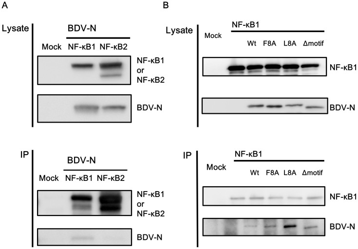
Figure 4. Interaction of BDV-N with NF-κB1.
(A) Lysates from cells expressing HA-tagged BDV-N were mixed with lysates from cells transfected with FLAG-tagged NF-κB1 or FLAG-tagged NF-κB2 for 30 min at 4°C. Mixed cell lysates were then immunoprecipitated with an anti-FLAG M2 monoclonal antibody using Protein G Dynabeads. SDS-PAGE and western blotting with an anti-HA monoclonal antibody and anti-FLAG M2 monoclonal antibody were performed. Full-length blots are presented in Supplementary Figure 3. (B) Lysates from cells expressing HA-tagged BDV-N or HA-tagged IPBN-alanine substitution in the first 8 (F8A) or last 8 (L8A) amino acids or -deletion mutants (Δmotif) of BDV-N were mixed with lysates from cells and transfected with the FLAG-tagged NF-κB1 for 30 min at 4°C. The mixed cell lysates were then subjected to an IP assay with anti-FLAG M2 monoclonal antibody using Protein G Dynabeads. Full-length blots are presented in Supplementary Figure 4.

