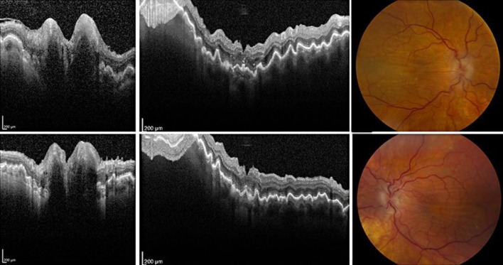
Fig. 1.
OCT and fundus photographs showing hypotony maculopathy bilaterally. OCT of the optic nerve head shows edema of the right eye (upper left) and left eye (bottom left). Vertical sections through macula OCT highlight the retinal folds (upper middle: right eye; lower middle: left eye). Color fundus photographs demonstrate similar findings (upper right: right eye; bottom right: left eye).

