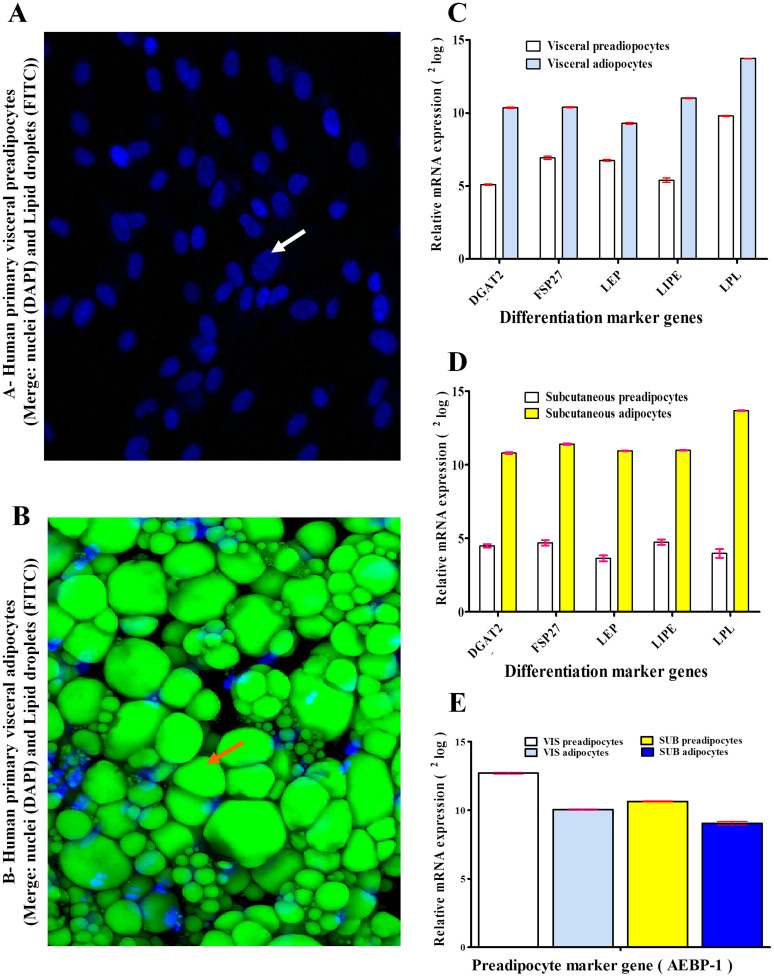
Figure 1.
(A–E). Differentiation of human visceral preadipocytes to adipocytes. (Panel A) shows human pre-adipocytes (no lipid droplets) and (Panel B) exhibits adipocytes (with medium-sized or single large lipid droplets). In panel A, nuclei and lipid droplets were stained with DAPI and FITC respectively. Nuclei were indicated by white arrow. In panel B, lipid droplets were stained with FITC and indicated by orange arrow. Almost the whole space of adipocytes is occupied by lipid droplets. Immune fluorescent confocal laser scan microscopy (IFCLSM) was applied to detect DAPI and FITC. (Panel C and D) display 5 differentiation marker genes for human primary visceral and subcutaneous adipocytes respectively. mRNA expression was expressed as2log (for example: the difference between human subcutaneous preadipocytes and adipocytes regarding leptin (LEP) expression is approximately 7, thus true difference is 27 ( = 128-fold) and was shown in y-as. Diacylglycerol O-acyltransferase 2 (DGAT2), hormone-sensitive lipase (LIPE), lipoprotein lipase (LPL) and fat specific protein-27 (FSP27). (Panel E) shows marker gene for human primary visceral and subcutaneous preadipocytes. Adipocyte enhancer-binding protein 1 (AEBP-1) was higher in human preadipocytes than human adipocytes.

