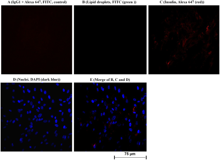
Figure 4.
(A–E). Confocal microscopy analysis was applied to detect and localize insulin protein in the human primary visceral preadipocytes. The cultured preadipocytes in six-well plates were incubated with IgG1 isotype and the detection antibody Alexa 647 (A; considered as negative control), lipid droplets were stained with FITC (B; green color); adipocyte was incubated with monoclonal antibody against human insulin. Bound antibodies were detected with Alexa 647 coupled goat anti-mouse (C; red color); preadipocytes were stained with DAPI to detect nuclei (D, blue color). E (merge) exhibits a merge of all three labeling; B (lipid droplets), C (insulin) and D (nuclei). Insulin protein was located in cytoplasm and on the surface of plasma membrane (C and E) of human visceral preadipocytes. Fluorescent labeling was used for all detections. Original magnification used was 400 times. The same results were found for human subcutaneous preadipocytes (Figure 3 A–G, SI).

