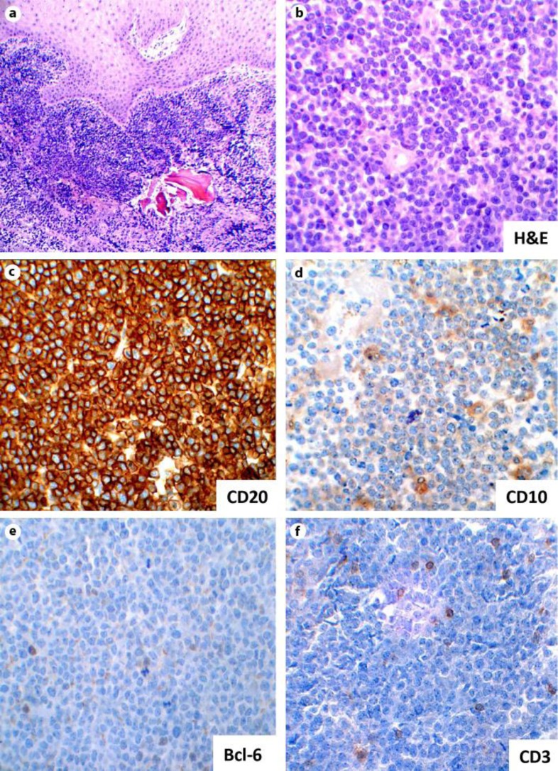
Fig. 2.
Right posterior mandible biopsy. a Photomicrograph showing multiple fragments of a squamous mucosa, soft tissue and bone with a dense lymphoid infiltrate (×10 magnification). b Photomicrograph showing an aggregate of atypical lymphoid cells (H&E staining; ×40 magnification). c Photomicrograph of immunohistochemical stain showing sheets of large mononuclear lymphoid cells positive for CD20 (×40 magnification). d Immunostaining for a CD10 showing a weak positivity (×40 magnification). e, f Scattered positivity is showing for CD3 and Bcl-6 immunostains (×40 magnification).

